Tubakia Leaf Blotch
Pathogen
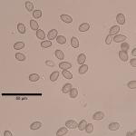
Tubakia leaf blotch is primarily attributed to the actions of Tubakia dryina. However, research has shown that at least six species of Tubakia are known to occur on Quercus in the eastern U.S., indicating that the disease is caused by a complex of Tubakia species (Harrington and McNew 2018).
Hosts
Numerous native and non-native oaks (Quercus spp.) serve as the primary hosts in the region. Secondary hosts include: chestnut (Castanea), hickory (Carya), maple (Acer), ash (Fraxinus) and beech (Fagus).
Symptoms & Disease Cycle
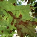
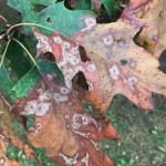
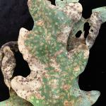
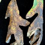
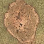
Tubakia leaf blotch is often a mid- to late-summer disease in southern New England, with symptoms first appearing in early to mid-July. Symptoms initiate as water-soaked, circular lesions that dry out and become reddish-brown to dark brown over time. The leaf spots can coalesce to create necrotic patches that consume large percentages of the leaf surface. It is not known whether a secondary infection cycle exists after the initial infection, but new lesions can develop well into August. Tubakia species are known endophytes, meaning they can colonize host tissues without causing noticable symptoms of disease. Therefore, new infections most likely initiate in the spring but disease symptoms take several weeks or months to develop. In addition to colonizing the foliage, Tubakia species can also colonize twigs but is not known as a cankering pathogen. The pathogen may overwinter within the canopy of infected oaks on leaves and petioles that did not properly abscise or within dead twigs. This allows the fungus to readily sporulate and infect newly developing tissues during the spring, when they are most susceptible to infection. In some cases, moderate defoliation can occur when numerous lesions form on infected leaves. However, typically the disease is not a serious threat to the overall health of infected oaks because the symptoms develop later in the growing season, when growth is mostly complete for the season. Therefore, only minor growth losses are expected from most outbreaks.
Management
Remove and discard fallen leaves to reduce inoculum at the site. Even if leaves (with petioles) are shed prior to new tissue development in the spring, they may be nearby, having avoided the autumn clean-up that removed downed foliage from most deciduous hardwoods. Therefore, a spring clean-up may be warranted where the disease is present. Tubakia species are native and widespread in the enviroment, therefore eradication is not possible. Chemical management is often unnecessary, but several fungicides are labeled for use against Tubakia and include: copper hydroxide, copper sulfate, mancozeb, myclobutanil, propiconazole, triadimefon+trifloxystrobin, trifloxystrobin and thiophanate-methyl. Despite the fact that symptoms do not develop until later in the growing season, infections are believed to initiate in the spring. Therefore, chemical application should take place as soon as new shoot development begins and on regular intervals, should wet conditions that are conducive to spore production persist.
Citation
Harrington T.C. and D.L. McNew. 2018. A re-evaluation of Tubakia, including three new species on Quercus and six new combinations. Antonie van Leeuwenhoek 111, 1003–1022. https://doi.org/10.1007/s10482-017-1001-9
